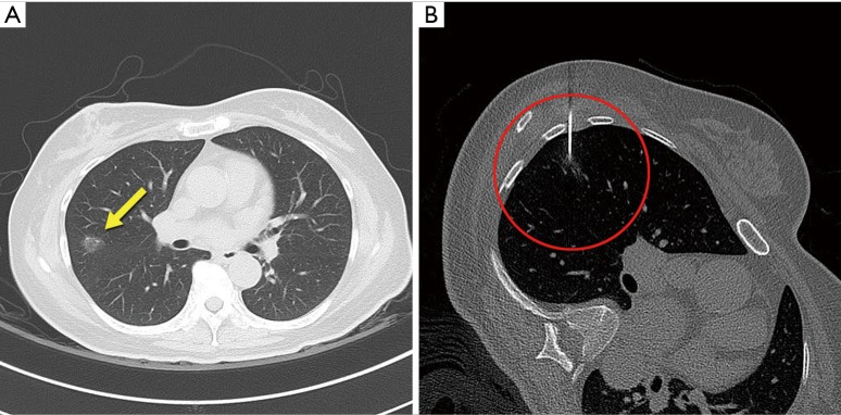
Figure 1.
CT images of GGO pulmonary nodule and CT-guided localization technique. (A) Chest tomography shows peripherally located ground glass opacity pulmonary nodule of the right upper lobe (axial view, arrow); (B) CT-guided percutaneous localization and injection of the mixture of lipiodol and India ink solution using 18-gauge Chiba needle. GGO, ground-glass opacity.

