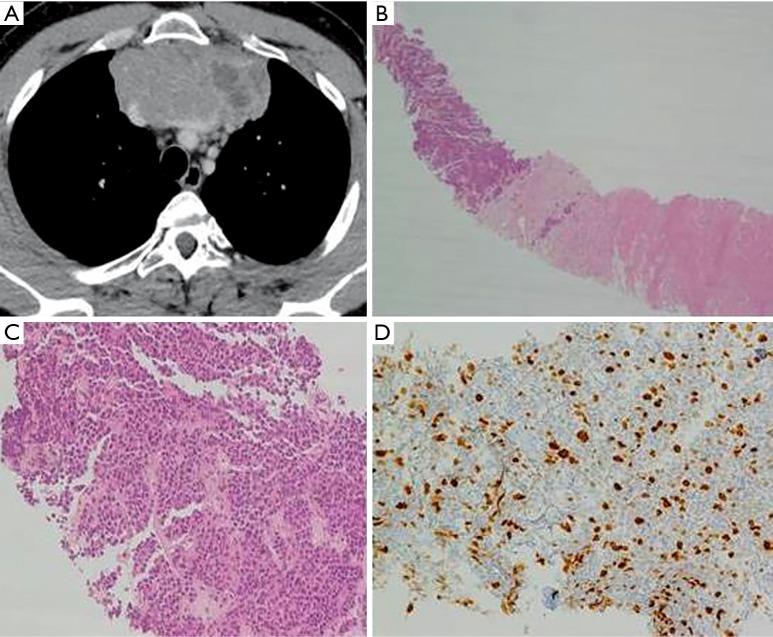
Figure 1.
(A) Chest CT-scan (before chemotherapy) showing a large tumor mass in the anterosuperior mediastinum invading the superior vena cava and the left anonymous vein; (B) biopsy sample (before chemotherapy) showing tumor cells in the left upper corner; connective and fibrotic tissue was present in the middle of the picture and a marked necrosis was observed in the right lower corner (EE ×4); (C) tumor tissue (before chemotherapy) showing clusters of medium-sized cells, eosinophilic cytoplasm with nuclear pleomorphism and small nucleoli arranged in trabeculae and immersed in a hyaline stroma with areas of necrosis (EE ×20); (D) biopsy tissue (before chemotherapy) showing a Ki67=20% (magnification ×10).

