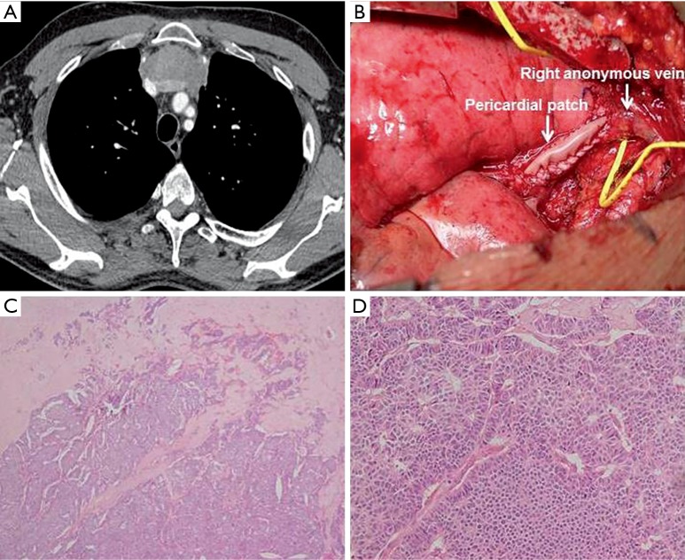
Figure 2.
(A) Chest CT-scan (after 4 cycles of neoadjuvant chemotherapy) showing a partial response with a significant reduction of the tumor mass. The left anonymous vein appears invaded by the tumor mass all along its mediastinal course; (B) intraoperative view showing the pericardial patch used to reconstruct part of the superior vena cava previously resected; (C) tumor tissue (after neoadjuvant chemotherapy) showing tumor cells and necrosis (EE ×4); (D) tumor tissue (after neoadjuvant chemotherapy)showing medium-size cells with nuclear pleomorphism and small nucleoli, arranged in trabeculae, nests and lobules (EE ×20).

