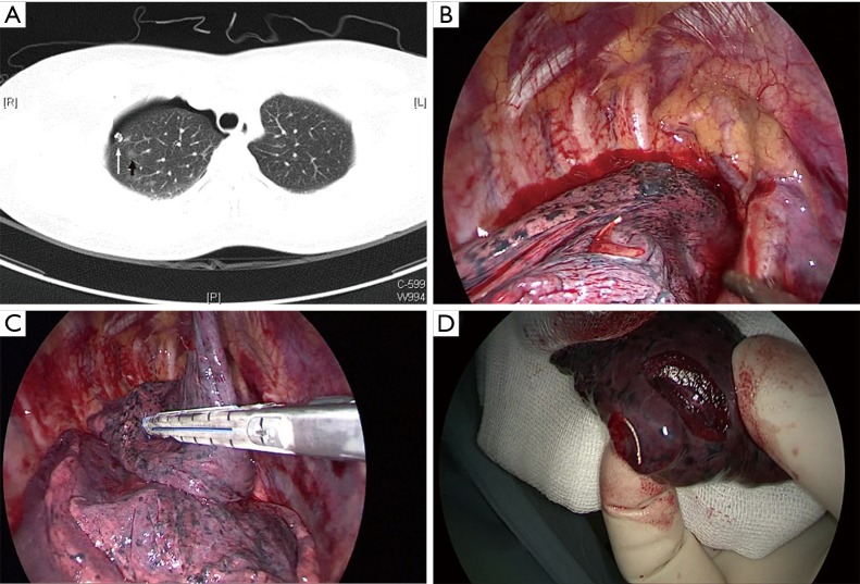
Figure 1.
A 65-year-old female patient with a 7-mm GGN situated in the RUL underwent the CT-guided microcoil marking procedure preoperatively and wedge resection by VATS. Histopathological analysis indicated AIS. (A) After induced minor pneumothorax, a microcoil was placed next to the lesion (white arrow, the marking microcoil; black arrow, the target lesion), shown by CT scan; (B) the tail of the microcoil was spotted on the visceral pleura surface during the VATS; (C) a VATS wedge resection was performed to remove the target lesion, the microcoil and the surrounding lung parenchyma; (D) inspection of the specimen confirmed the successful resection of the lesion and the marking microcoil. GGN, ground-glass nodule; CT, computed tomography; VATS, video-assisted thoracoscopic surgery; AIS, adenocarcinoma in situ; RUL, right upper lobe.

