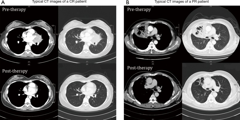
Figure 2.
Typical CT images of a patient obtaining CR (A) and a patient with PR (B). In patient A who have achieved CR after received six cycles of rituximab and cladribine, a post-therapy CT scan 1 month after treatment shows complete resolution of former lesion near the left-lung hilus. Patient B was documented as PR in post-treatment evaluation CT scan and had continuous improvement during follow-up. CT scan performed 1 year after treatment shows shrinkage of the former mass near right-lobar and residual fibrous stripes with dilated bronchi. CT, computed tomography; CR, complete remission; PR, partial remission.

