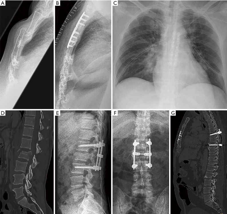
Figure 4.
Radiography of the initial injury and after operative treatment. (A) Dislocation of angulus sterni; (B) anatomical reduction after anterior plating; (C) chest X-ray postoperative shows two sternal plates and chest tubes bilateral; (D) unstable fracture of L2; (E) internal fixator L1-L3 lateral view; (F) anterior-posterior view; (G) the alignment of the trunk has been restored through a combined stabilization of the sternum and the spinal column.

