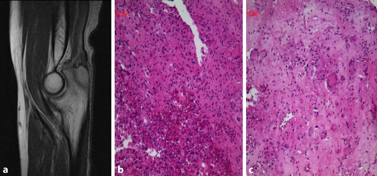
Fig. 2.
a MRI of right elbow revealed a slightly long T1, long T2 signal of a size of 7 × 0.9 cm in the rear end of right olecranon and its partial edge was hazy. b, c The histological findings showed typical epithelioid granuloma in a background of marked inflammation comprising of sheets of neutrophils, histiocytes, plasma cells, and increased lymphocytic infiltration. Specific stains showed acid-fast staining (+) and periodic acid Schiff stain (PAS) (−) (magnification, ×200)

