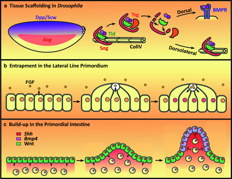
Fig. 2.

Influence of tissue architecture on signaling range. a Dorsally secreted Dpp–Scw heterodimers bind to collagen IV along with Sog and Tld. Inhibitory complex formation on Collagen IV (ColIV) acts to restrict dorsolateral signaling but also enables release of a freely diffusing shuttling complex by Tsg. Proteolytic cleavage of Sog by Tld releases the Dpp–Scw heterodimer which, if dorsally localized, can promote Dpp signaling. However, if cleavage occurs dorsolaterally, then the inhibitory complex is reformed. b Left panel the zebrafish lateral line epithelial primordium migrates along the flanks of the fish and secretes FGF ligands. Middle apical constriction of the epithelial cells drives the formation of a ‘rosette’ shaped organ with a central microlumen into which FGF is secreted. Right after FGF levels reach a threshold, the cells stop migrating and continue mechanosensory organ development. c Left panel the early chick gut epithelium (green) retains a stem cell-like identity (Lgr+, Sox9+) in response to Wnt signaling and secretes Shh to drive low-level Bmp4 expression in the underlying mesenchyme. Middle mechanical stress later results in the buckling of the epithelium. Right the concentration of Shh at the tip of the folds results in increased Bmp4 expression in the fold mesenchyme. BMP4 signals back to the gut epithelium to repress Wnt signaling to promote the differentiation of the fold epithelium, or villus, and restrict the stem cell pool to those cells at the base of the fold. For simplicity, the cytoplasmic colour is used to depict the stem cell or differentiated fates, depending on the presence or absence of Wnt signaling, respectively, whereas the nuclear colour depicts expression of Shh or Bmp4
