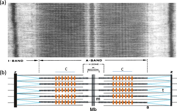
Fig. 1.
a Electron micrograph of a longitudinal section from frog muscle showing the A-band where the bipolar myosin filaments are (see b). The actin filaments run through the I-band from the Z-line and into the A-band where they interdigitate with the myosin filaments. The actin filaments end at the edge if the H-zone. b Schematic diagram of the overlapping myosin (m) and actin (a) filaments. The protein titin (t; blue) runs from the M-band (Mb) along the myosin filaments and then across the I-band to the Z-line (Z). C-protein (MyBP-C) forms a set of stripes in the two halves of the A-band at the positions marked C (orange stripes). C-protein binds to the myosin filament backbone and in some conditions extends out to bind to actin. Figure modified from Fig. 5 of Squire et al. (2005)

