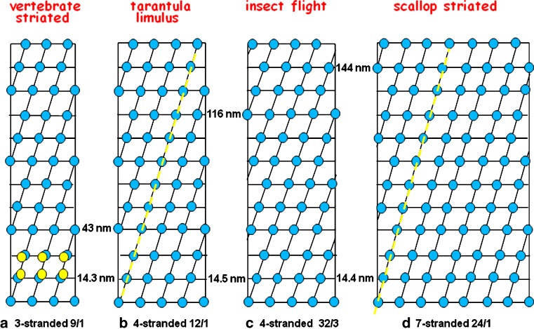
Fig. 5.
The accepted symmetries for the myosin filaments in a vertebrate striated muscle, b tarantula and Limulus myosin filaments, c insect flight muscle (Lethocerus) and d scallop striated adductor muscle. These structures all have the myosin head pairs grouped in very similar surface arrays; a common axial repeat of about 14.3–14.5 nm and a common lateral spacing of around 10–15 nm). The slightly different tilt angles of the helical strands give rise to the different observed long repeats. The yellow dashed lines in (b) and (d) indicate the long-pitched helices along which the Class III head interactions (Fig. 6) were originally thought to occur

