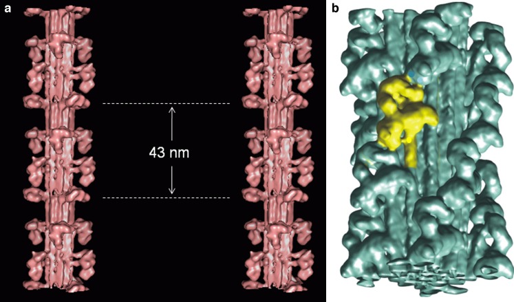
Fig. 7.
Recent images of myosin filament models from a vertebrate striated muscle determined by modelling the low-angle X-ray diffraction pattern from bony fish muscle and shown in stereo (AL-Khayat and Squire 2006) with the backbone shown as a molecular crystal structure (Squire 1973; Chew and Squire 1995), and b tarantula muscle determined by single particle analysis of filaments viewed frozen-hydrated (Woodhead et al. 2005; the image is inverted from the original to make it consistent with all other figures in the review (M-band towards the bottom). In both cases, the heads are mostly in the Wendt-like pairing in which the two heads of one molecule interact in a parallel fashion (Class 1 in Fig. 6). The exception is the crown in vertebrate striated muscle filaments at the dotted lines in (a) which appears very different

