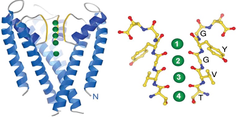
Fig. 3.
Crystal structure of the KcsA potassium channel (Doyle et al. 1998) showing the selectivity filter (yellow) and binding sites for K+ ions (green). It has a tetrameric structure but only two of the monomers are shown for clarity. A magnified view of the filter depicting the carbonyl oxygens (red) that form the four binding sites is shown on the right. A fifth binding site (S0) is formed just above the S1 site

