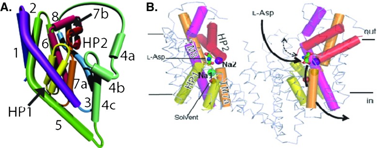
Fig. 5.
The topology of a single monomer of GltPh (left). Two monomers of GltPh are shown in the membrane (right). TM1–6 are in ribbon representation, TM7, 8, and HP1, HP2 are shown as cylinders, and bound aspartate is shown in stick representation. HP2 serves as an extracellular gate and controls access of Asp to its binding site. Na+ ion #1 is below the binding site, while Na+ ion #2 is above it and serves as a lock on this gate

