Figure 2.
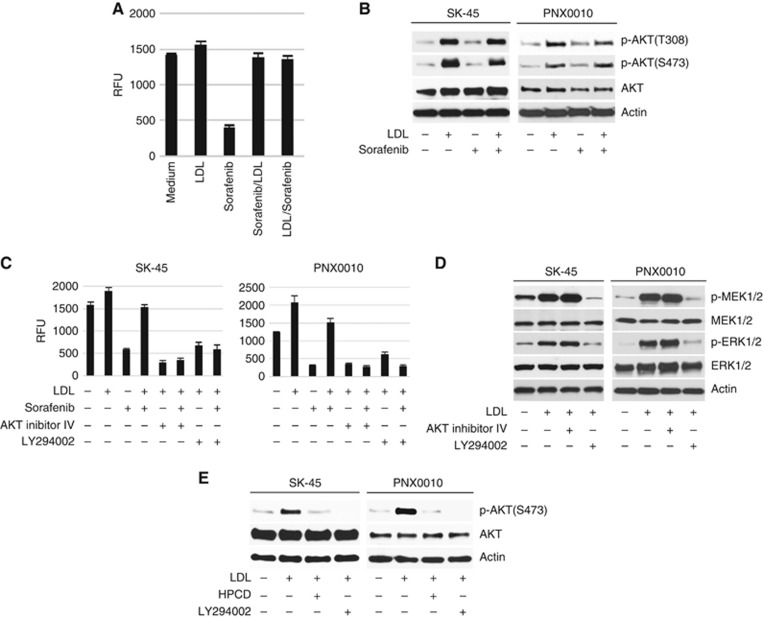
Inhibition of PI3K/AKT signalling enhances the antitumour efficacy of TKIs in the presence of LDL.(A) SK-45 cells were pre-treated with sorafenib (1 μM) for 3 h followed by the addition of LDL (100 μg ml−1), or in the reverse order. Cell viability was analysed by CellTiter Blue assay. (B) Sorafenib does not affect LDL-induced activation of AKT. SK-45 cells were pre-treated with sorafenib (2.5 μM) for 1 h followed by the addition of LDL (100 μg ml−1) for 1 h. (C) SK-45 and PNX0010 cells were pre-incubated with AKT inhibitor IV (1 μM) or PI3K inhibitor LY294002 (10 μM) for 1 h followed by incubation with sorafenib (1 μM) and/or LDL (100 μg ml−1) for 72 h. Cell viability was analysed by CellTiter Blue assay. (D) The addition of LDL activates MEK/ERK signalling. SK-45 and PNX0010 cells were pre-incubated with AKT inhibitor IV (1 μM) or LY294002 (10 μM) followed by the addition of LDL (100 μg ml−1) for 1 h. (E) The effect of HPCD on LDL-mediated AKT activation in RCC cells. SK-45 and PNX0010 cells were pre-treated with LDL (100 μg ml−1) for 1 h followed by the addition of HPCD (10 mM) for 2 h. LY294002 (10 μM) was used as a control in this experiment.

