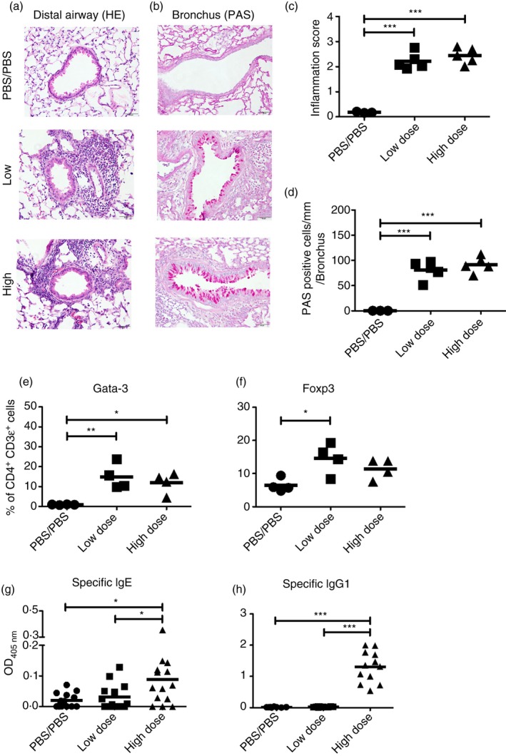
Figure 2.

Low‐dose and high‐dose priming elicit comparable inflammatory cell infiltration around airways and vessels and airway mucus secretion and similar GATA‐3 and Foxp3 expression, but low‐dose priming does not produce a significant IgE or IgG1 response. Representative images for (a) haematoxylin and eosin (H&E) and (b) periodic acid‐Schiff's (PAS) stained lung tissue sections are shown. Magnification × 400. Scale black bar; 50 μm. (c) Scoring of H&E‐stained lung sections for severity of inflammation. The sections scored blind by using a five‐point scoring system as follows, 0, normal; 1, few cells; 2, a ring of inflammatory cells one‐cell‐layer deep; 3, a ring of inflammatory cells two to four cell‐layers deep; 4, a ring of inflammatory cells more than four cell‐layers deep. Fifty airways and blood vessels were scored per lung. (d) Scoring of PAS‐stained lung section for mucus‐secreting cells on airways. The lung sections were scored blind, using five points along 200 μm of bronchi per sample. In central airway, five spots per lung were randomly selected, 200 μm was measured along the bronchi, and PAS‐positive cells were counted. The score was determined as the sum of cells expressed as the number of PAS‐positive cells per unit length (mm) of basement membrane on bronchus (n = 5). (e) The expression of GATA‐3 and (f) Foxp3 in lung cells 1 day after the last challenge. Numbers indicate the percentage of positive CD4+ CD3ε+ (n = 4). (g) Blomia tropicalis extract (BTE) ‐specific IgE and (h) IgG1 levels in serum. Data are pooled from four independent experiments with three to four mice/group/experiment (n = 8−14). Statistical analysis was performed using analysis of variance with Tukey's test, *P < 0·05, **P < 0·01, ***P < 0·001.
