Figure 3.

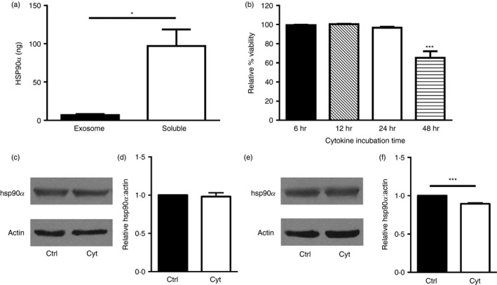
Mechanisms of heat‐shock protein 90α (hsp90α) release. Human βLox5 cells were treated with pro‐inflammatory cytokines and after 24 hr the extracellular medium was collected and fractionated to separate exosomes and the residual soluble media fraction, for analysis of hsp90α distribution. Total hsp90α levels in each fraction were determined by ELISA (a). βLox5 cells were treated with or without pro‐inflammatory cytokines for various times, and cellular release of cytoplasmic lactate dehydrogenase was monitored for up to 48 hr (b). Human beta cells βLox5 (c, d) and 1.1B4 (e, f) were treated for 24 hr with media alone (Ctrl) or interleukin‐1β (IL‐1β), tumour necrosis factor‐α (TNF‐α) and interferon‐γ (IFN‐γ) (Cyt). Total cell hsp90α and actin protein levels in each sample were assessed by immunoblotting. (c, e) Representative images of Western blots, (d, f) densitometry results indicating relative cellular hsp90 levels compared with actin expression from n = 3 experiments. Data are mean + SEM of n = 3 experiments. *P < 0·05, ***P < 0·001.
