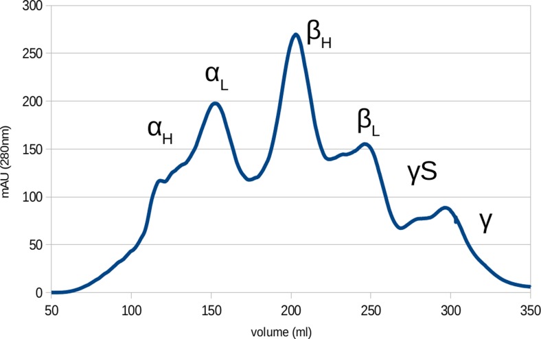
Fig. 5.
Chromatogram of lens fibre cells cytoplasm from bovine eye lens separated with gel filtration chromatography. Significant protein groups are marked. α- and β-crystallins separate into high and low mass elution peaks corresponding to different tertiary structures and γS-crystallin elutes separately to the other γ-crystallins

