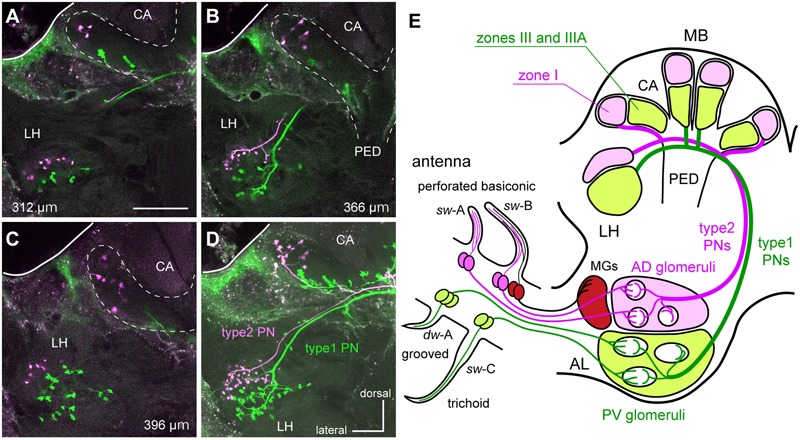
FIGURE 2.

The segregated parallel pathways from the periphery to higher brain centers in the cockroach brain. (A–D) A type1 PN (green) and a type2 PN (magenta) differentially labeled in the protocerebrum. Serial optical sections (A–C) and a stacked image (D) reveal that terminal regions of type1 PN are segregated from those of the type2 PN in the MB calyces (CA) and the LH. Depths of the serial optical images from anterior to posterior are indicated in each of panels (A–C). Twenty-five serial optical images obtained every 4-μm are stacked in (D). Bar in (A) = 100 μm. (E) Schematic drawing of parallel pathways from the periphery to higher brain center in the cockroach brain. OSNs in perforated basiconic sensilla (single-walled A [sw-A] and sw-B) selectively terminate in antero-dorsal group glomeruli (AD glomeruli), whereas those in trichoid sensilla (sw-C) and grooved basiconic sensilla (double-walled A [dw-A]) projects to the postero-ventral group glomeruli (PV glomeruli: Watanabe et al., 2012b). Therefore, type1 PNs with dendrites in PV glomeruli and type2 PNs with dendrites in AD glomeruli form two segregated parallel pathways from the periphery to higher brain centers. Nomenclatures of MB calyces were described in previous articles (Mizunami et al., 1998; Strausfeld and Li, 1999a,b). MGs, macroglomeruli; PED, pedunculus.
