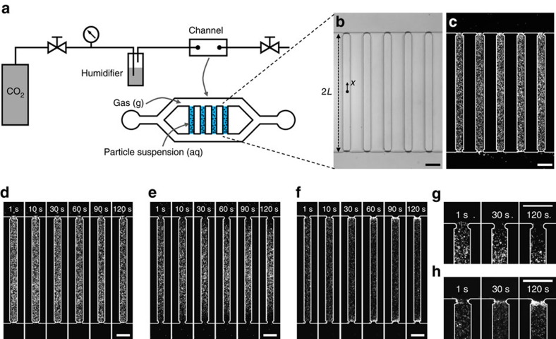
Figure 1. Motion of colloidal particles upon exposure of aqueous suspensions to gas.
(a) Experimental set-up for creating a liquid column with a stable gas–liquid interface. (b) Bright-field and (c) corresponding fluorescence microscopy images of suspension-filled channels exposed to gas, where the length of the channel is 2L=800 μm. The width and the height of the channels are 60 and 40 μm, respectively. The ends of the pores are slightly contracted to effectively fix the liquid columns. (d) Image sequence showing particles (polystyrene, diameter 0.5 μm) exposed to N2. (e,f) Image sequences showing directed motion of particles with (e) negative (polystyrene, diameter 0.5 μm) and (f) positive (amine-functionalized polystyrene, diameter 1 μm) surface charge exposed to CO2. (g,h) Close-up images of the time evolution of the (g) negatively charged and (h) positively charged particles near the gas–liquid interface during exposure to CO2. The gas pressures in all cases are 136 kPa. All scale bars are 100 μm.

