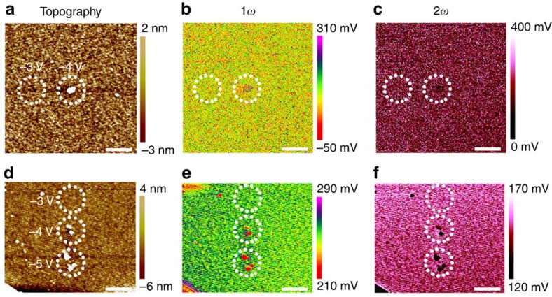
Figure 3. Electrostatic force microscopy characterization after negative voltage sweeps.
(a–c) Topographic (a), 1ω (b) and 2ω (c) measurements on the HfO2/TiN sample undergoing negative voltage sweepings in preceding C-AFM measurements. The voltage amplitudes were –3 and –4 V at different locations. Scale bar, 1 μm. (d–f) Topographic (d), 1ω (e) and 2ω (f) measurements on the HfO2/TiN sample undergoing negative voltage sweepings. The voltage amplitudes were –3, –4 and –5 V at different locations. Scale bar, 1 μm. The regions of interest are marked by circles. These repeated studies showed highly consistent results, where no charge accumulations were observed in case of negative voltage sweeps until structural deformations took place.

