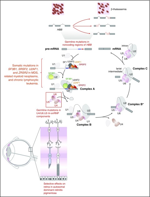
Figure 1.
Diagram of the complexes involved in RNA splicing and how trans-acting mutations in splicing factors as well as mutations in sites required for splicing of genes in cis may result in tissue-specific phenotypic effects. The individual steps and components of each spliceosomal complex have been described recently in other reviews.58-62 Germ line mutations in the genes encoding 6 different members of the U4/U6.U5 tri-small nuclear ribonucleoprotein particle (tri-snRNP; red box) result in the retinal degenerative disorder known as autosomal dominant retinitis pigmentosa. Despite the ubiquitous expression of these proteins and their role in core RNA splicing function required in every cell, overt phenotypic effects of these mutations are only apparent within the retina. In contrast to the U4/U6.U5 tri-snRNP mutations in autosomal dominant retinitis pigmentosa, mutations in the RNA splicing factors SF3B1, U2AF1, SRSF2, and ZRSR2 are enriched in leukemias and subsets of epithelial malignancies. Again here, how mutations in core RNA splicing factors expressed in numerous cell types are enriched in diseases of specific lineages remains to be addressed. In addition to mutations in RNA splicing proteins, mutations in coding or noncoding regions are required for RNA splicing of a gene in cis. The earliest examples of such an alteration are the mutations within HBB that are well known to be associated with β-thalassemia. Despite the fact that such mutations may occur in the germ line, the direct phenotypic effects of these mutations are specific to the hematopoietic system given the importance of hemoglobin β to red blood cell function.

