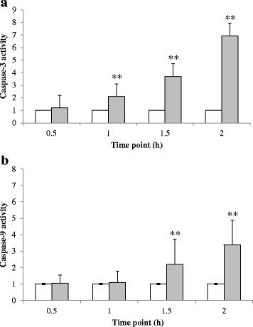
Fig. 3.

Effect of cardol treatment at 14 μg/mL on the activity of (a) caspase-3 and (b) caspase-9 in SW620 cells. White bars represent the control and grey bars are the cardol-treated cells. Data are expressed as the mean ± SE, derived from three independent repeats. Compared to the control, ** represent a significant difference at the p < 0.01 level
