
Fig. 2.
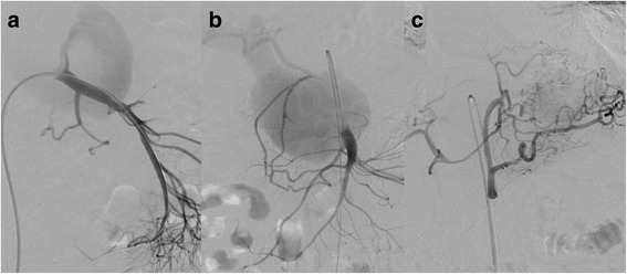
Preoperative angiography showing a pseudoaneurysm of the proximal superior mesenteric artery (a, b) with fistulization to the portal vein (b). The pseudoaneurysm is filled partially via a large defect in the top of the superior mesenteric artery (a). There is no connection between the common hepatic artery and the coeliac trunk (c)