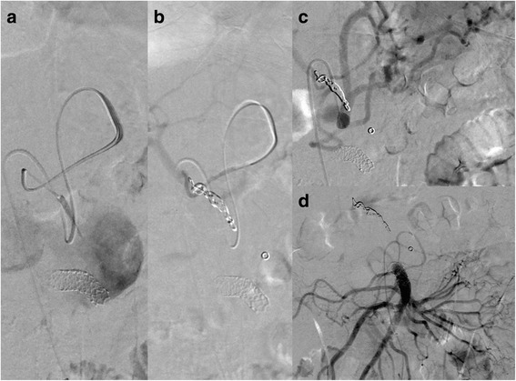
Fig. 3.

Postoperative angiography showing a covered stent (Bentley Begraft® diameter 8 mm, length 37 mm) covering the defect in the superior mesenteric artery and refilling of the pseudoaneurysm via the common hepatic artery (a). Embolization of the common hepatic artery with multiple Hilal microcoils (3, 4, and 5 mm wide and 3 and 6 cm long) after selective catheterization of the common hepatic artery via the left and then the right gastric artery (a, b). Complete exclusion of the pseudoaneurysm sac after coiling of the common hepatic artery and covered stenting of the superior mesenteric artery (c, d)
