Figure 5.

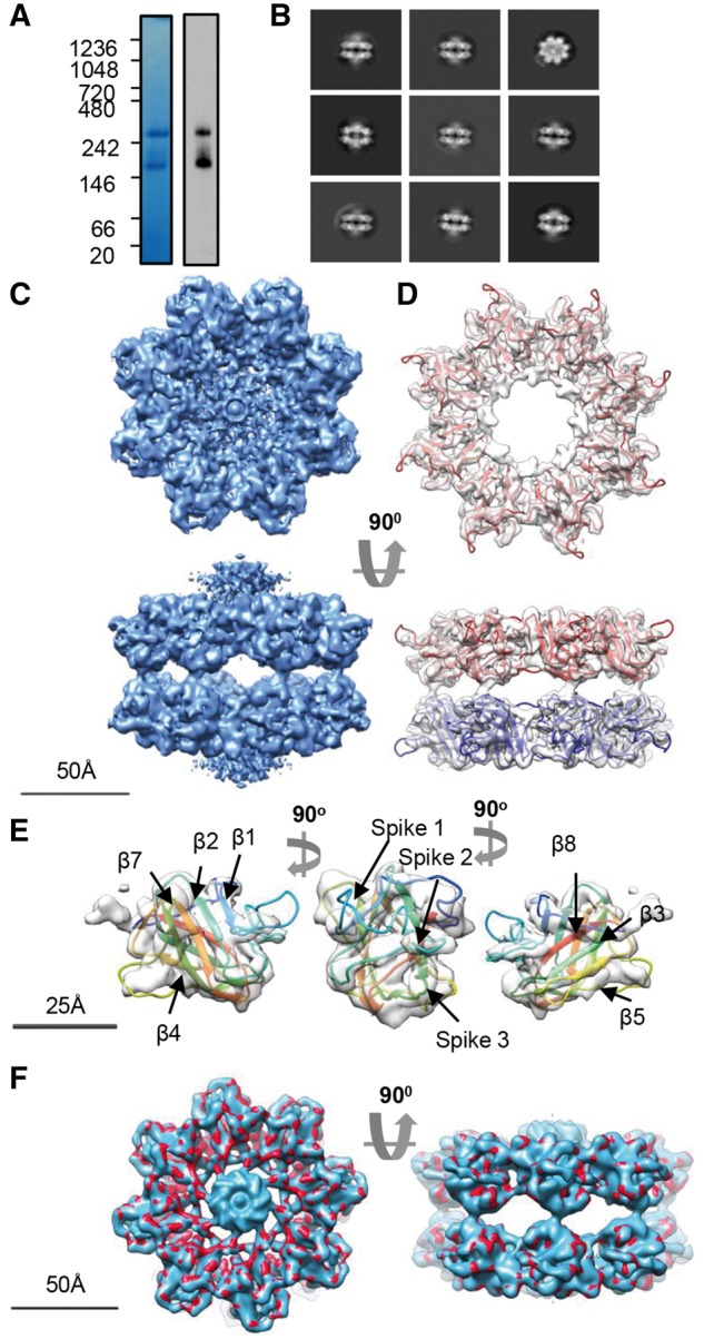
Cryo-EM analysis of the R141H dimer of octamers. (A) Blue-native PAGE and native-blot analysis of R141H retinoschisin. (B) Reference-free class averages of R141H dimer of octamers. Box size = 33 nm (C) Three-dimensional reconstruction at 4.2Å resolution of the R141H dimer of octamers. Shown is the unmasked map. (D) Fitting of the R141H discoidin domain model (residues 63-219) shown in red, with associated subunits in the opposing octamer shown in blue. The central density of Rs1 domains has been removed for clarity. (E) Fit into single subunit showing the fitting of the β-sheets and positions of the spike regions. (F) Comparison between wild-type hexadecamer (blue, emd_6425) and R141H retinoschisin (red) structures at 5Å resolution.
