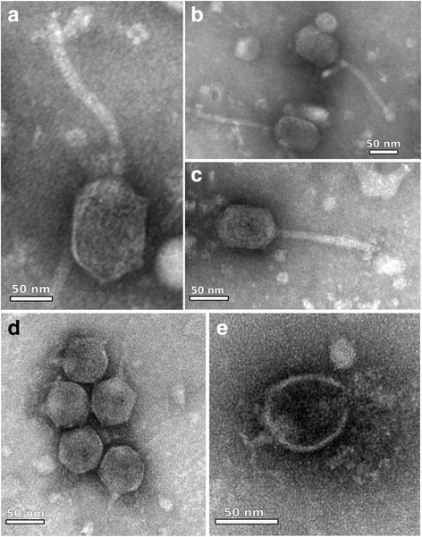
Fig. 5.

Electron micrographs of phages ZC01, ZC03 and ZC08. Transmission electron micrographs of negatively stained Pseudomonas phages virions found in composting: a–c Pseudomonas phage ZC01 with typical morphology of members of the Siphoviridae family; (d, e) Pseudomonas phages ZC03 and ZC08, full virions and empty shelled, respectively, with typical morphology of members of the Podoviridae family. Note the short tail
