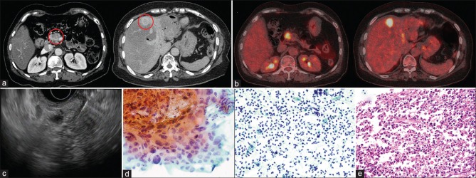
Figure 1.
(a) Computed tomography showing a pancreatic mass accompanied with a liver mass at segment 4. (b) Positron emission tomography original magnification ×40 showing hypermetabolic lesions in the pancreas and liver. (c) Endoscopic ultrasound-guided fine-needle aspiration concurrently performed in pancreas and liver. (d) Cytological examination showing adenocarcinoma of the pancreas (left). However, liver fine-needle aspiration showed only inflammatory cells (right). (e) The patient underwent pancreaticoduodenectomy. Surgical specimen of liver acquired during pancreaticoduodenectomy shows no malignant cells in liver

