Figure 4.

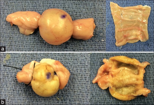
Ablation-induced macroscopic changes in the external (left panels) and internal (right panels) layers of 60°C (a) and 90°C (b) ablated cyst models showing color changes from pink to yellow as well as progressive dehydration of the tissue

Ablation-induced macroscopic changes in the external (left panels) and internal (right panels) layers of 60°C (a) and 90°C (b) ablated cyst models showing color changes from pink to yellow as well as progressive dehydration of the tissue