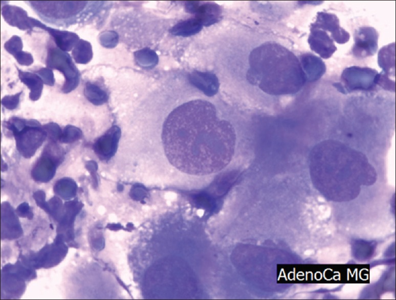
Figure 2.

Adenocarcinoma of the pancreas (May Grünwald Giemsa staining, ×400) Note that the nuclei are inhomogenous and reaching more than 2, 5 fold the size of erythrocytes (some are visible in the left upper end of the picture), mitotic figures are clearly to be seen in the cancerous cells
