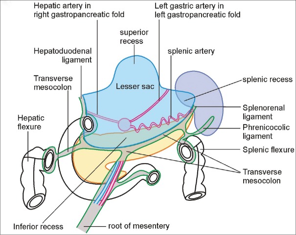
Figure 18.
A schematic diagram showing the lesser sac following the removal of the stomach and the transverse colon. The line of attachment of the transverse mesocolon and the mesentery is shown. The left gastric artery and common hepatic artery project into the lesser sac, raising the left and right gastropancreatic folds, which divide the lesser sac into the superior and inferior recesses. The splenic recess of the lesser sac is seen extending toward the splenic hilum

