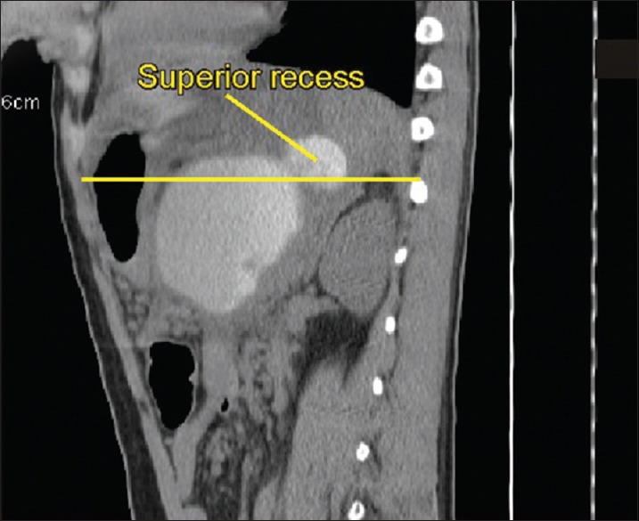
Figure 20.

A case of pancreatic duct leak into the lesser sac. The computerized tomography scan was performed after endoscopic retrograde cholangiopancreatography. The contrast is filling the lesser sac. The yellow line demarcates an approximate area above which the superior recess is seen
