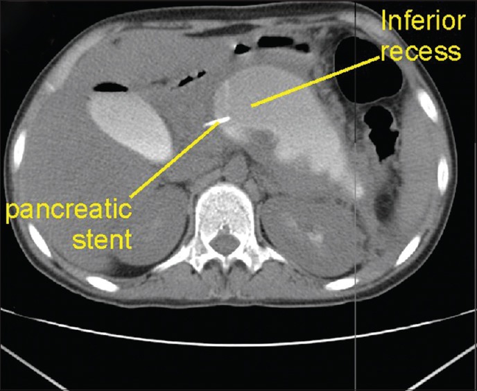
Figure 25.

A case of a pancreatic duct leak into the lesser sac. Computerized tomography scan was performed after endoscopic retrograde cholangiopancreatography. The contrast is filling the lesser sac. This patient had pancreatic duct disruption into the lesser sac and a pancreatic duct stenting was done. The stent is seen reaching up to the inferior recess of the lesser sac, which lies anterior to the upper pole of the left kidney. The presence of contrast within the recess is also noted
