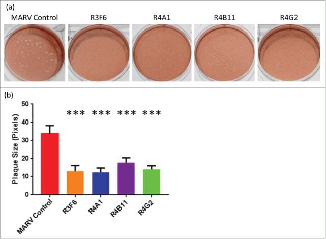
Figure 4.

(A) Photographic representation of viral plaques and (B) corresponding sizes in Vero E6 cells. ***All plaque sizes were highly significant to a p-value <0.0001 by utilizing a two-tailed t-test for the four antibody fragments tested against control virus.
