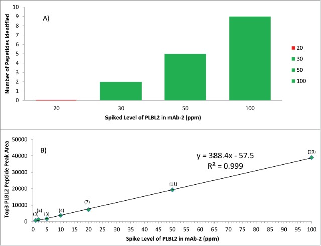
Figure 5.

Comparison of DDA (5A) and DIA SWATH MS (5B) methods to detect PLBL2 at different spiked levels in mAb2. Green represents successful detection, while red indicates failure to detect. The detection sensitivity of DDA is shown in 5A, and the sensitivity and linearity of DIA SWATH MS TOP 3 method is shown in 5B, with the number of peptide identified shown in parentheses. PLBL2 peak area measurement by SWATH (Top 3 peptides) at ≥ 5 ppm levels had observed RSD values ranging from 2.1–5.8%, based on triplicate injections.
