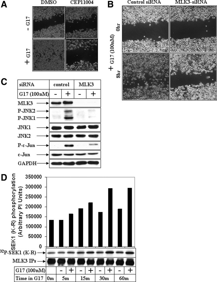
Fig. 5.
Effect of inhibition of MLK3 pathway on G17-induced JNK activation and migration. A, AGSE cells were treated with 100 nm G17 after a pretreatment with 1 μm pan-MLK inhibitor CEP11004. Wound-healing assays were performed next and pictures obtained as described in Fig. 1A. B, Subconfluent AGSE cells were transiently transfected with either 50 nm control-siRNA or 50 nm MLK3-siRNA, followed by wounding and G17 treatment as described in Fig. 4B. C, Subconfluent AGSE cells were transiently transfected with either control-siRNA or MLK3-siRNA, followed by G17 treatment for 30 min. Western blot analyses were performed with the antibodies indicated. D, Endogenous MLK3 immunoprecipitated from AGSE cells treated in the absence (−) or presence (+) of G17 for the indicated periods of time was subjected to in vitro kinase assay using recombinant SEK1 (K-R) as substrate and [γ32P]ATP. The samples were fractionated by SDS-PAGE and transferred to membranes, and the radiolabeled bands were detected by phosphoimaging screens. Western blots of the immunoprecipitates were shown in the bottom to indicate equal input. DMSO, Dimethylsulfoxide; GAPDH, glyceraldehyde-3-phosphate dehydrogenase; IPs, immunoprecipitations.

