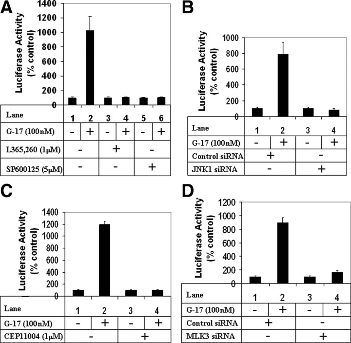
Fig. 6.
Role of MLK3 and JNK1 pathways on G17-induced MMP7 transcription. A, Subconfluent AGSE cells were transiently transfected with the MMP7-luciferase vector along with a β-Gal vector (for normalization of transfection). Forty-eight hours post-transfection, cells were treated with G17 after a pretreatment with either none (lanes 1 and 2), or with CCK2R antagonist L365,260 (lanes 3 and 4), or JNK inhibitor SP600125 (lanes 5 and 6), and luciferase and β-Gal assays were performed. The RLU/β-Gal values were represented as percent control, considering the corresponding untreated samples as control. B, AGSE cells were transfected as in panel A along with either control siRNA (lanes 1 and 2) or JNK1 siRNA (lanes 3 and 4). Luciferase and β-Gal assays were performed after G17 treatment as in panel A. C, Cells transfected with MMP7-luc and β-Gal vectors were treated with G17 after pretreatment with pan-MLK inhibitor CEP11004. Luciferase and β-Gal assays were performed next. D, AGSE cells were transfected with MMP7-luc or β-Gal vectors along with either control siRNA (lanes 1 and 2) or MLK3 siRNA (lanes 3 and 4), followed by G17 treatment. Luciferase and β-Gal assays were performed next. Each transfection (A–D) was performed in triplicate, and the data represent the mean ± sd of at least two independent experiments.

