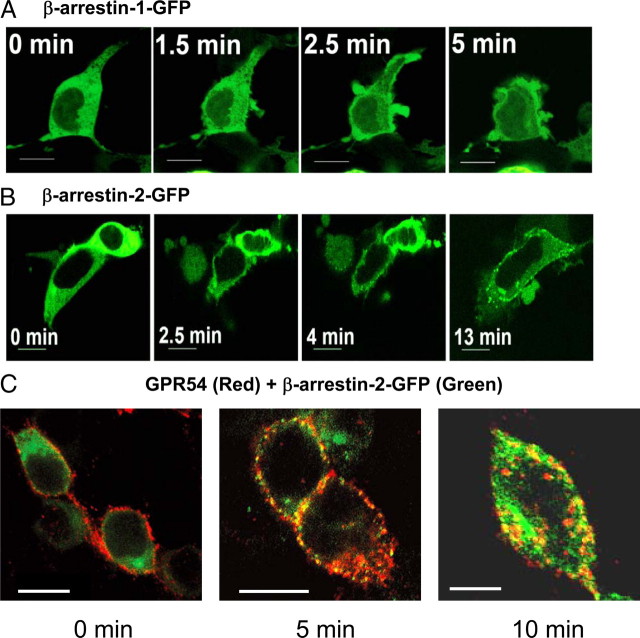
Fig. 4.
Activation of GPR54 stimulates the translocation of cytoplasmic β-arrestin-1 and -2 to the plasma membrane in HEK 293 cells. HEK 293 cells coexpressing FLAG-GPR54 and GFP-tagged β-arrestin-1 (A) or -2 (B) were stimulated with 100 nm Kp-10 and analyzed for the translocation of β-arrestin using live-cell confocal imaging. C, HEK 293 cells transfected with 10 μg FLAG-GPR54 and 5 μg β-arrestin-2-GFP were surface labeled at 4 C (0 min) using rabbit anti-FLAG antibody followed by agonist treatment (100 nm Kp-10) at 37 C for 5 or 10 min. After fixation, the localization of GPR54 was detected by Alexa Fluor 568 conjugated to antirabbit IgG and imaged by confocal microscopy. The overlay images show the localization of FLAG-GPR54 (red) and β-arrestin-2-GFP (green). Colocalization of both molecules is shown as yellow puncta. Scale bars, 10 μm.

