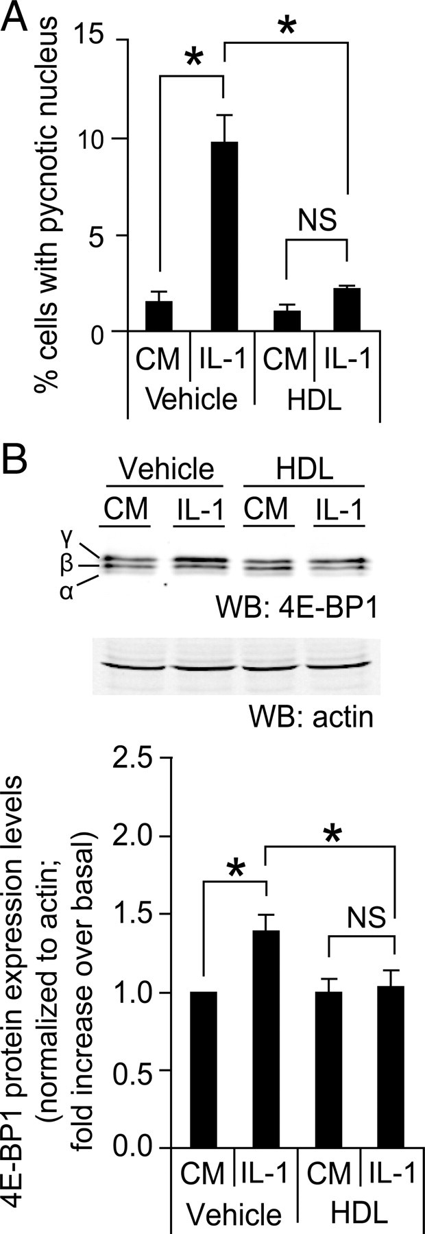
Fig. 4.

Effect of HDLs on IL-1β-induced apoptosis and increased 4E-BP1 levels. A, MIN6 cells were grown 2 d in complete medium, washed twice with PBS, and then incubated for 72 h, either in control conditions [culture medium containing serum (CM)] or treated with 10 ng/ml of IL-1β in the absence (vehicle) or in the presence of 1 mm HDLs. Apoptosis was then determined. The results correspond to the mean ± sem of four independent experiments. The threshold of significance was at P = 0.0167 (three comparisons). B, Alternatively, the cells treated or not with IL-1β were lysed and Western blot analysis was performed as described in Fig. 3B. The values for 4E-BP1 in the bar graph (black bars, mean ± sem; seven independent experiments) were normalized to actin levels. The threshold of significance was at P = 0.0167 (three comparisons). NS, Nonsignificant; WB, Western blot. *, Statistically significant as defined in Materials and Methods; α, hypophosphorylated form; β, intermediate phosphorylated form; γ, hyperphosphorylated form.
