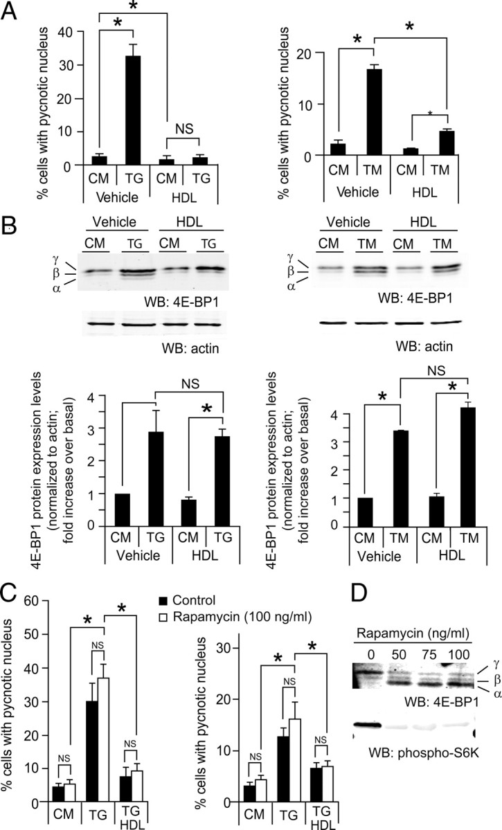
Fig. 6.

HDLs protect form ER stress-induced apoptosis independently of 4E-BP1. A and B, MIN6 cells were plated at 0.5 million per well in six-well plates and cultured for 2 d. They were then treated with 0.5 μm TG or 2 μg/ml TM in the presence or in the absence of 1 mm HDL for 24 h. Cells were then fixed, and apoptosis was determined by scoring the percentage of cells displaying pycnotic nucleus (panel A). Alternatively, the cells were lysed and Western blot analysis to assess 4E-BP1 expression was performed (panel B). The bar graphs in panel B represent total 4E-BP1 expression levels. The results of both panels correspond to the mean ± sem of three independent experiments. The threshold of significance was at P = 0.0167 (three comparisons). C, Cells were treated as in panel B but in the presence or in the absence of 100 ng/ml of rapamycin for 18 h. D, The functionality of rapamycin was checked by blotting for 4E-BP1 and phospho-S6K (panel D). The results of panel D correspond to the mean ± sem of four independent experiments. The threshold of significance was at P = 0.01 (five comparisons). CM, Complete medium; NS, nonsignificant; WB, Western blot. *, Statistically significant as defined in Materials and Methods; α, hypophosphorylated form; β, intermediate phosphorylated form; γ, hyperphosphorylated form.
