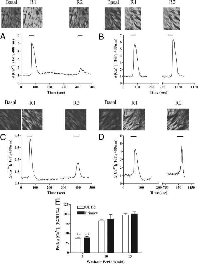
Fig. 3.
Desensitization and resensitization of OTR-stimulated Ca2+ signaling in ULTR and primary myometrial cells. Cells loaded with the Ca2+-sensitive dye Fluo4 were subjected to the OTR desensitization protocol, where cells were stimulated with oxytocin (R1, 100 nm, 30 sec) followed by variable washout periods and rechallenge with oxytocin (R2, 100 nm, 30 sec). Representative images and traces indicate the extent of OTR desensitization 5 min (A and C) and 15 min (B and D) after initial oxytocin challenge in ULTR (A and B) and primary myometrial (C and D) cells. OTR desensitization was determined as the relative change in R2 response compared with R1. Cumulative data (E) are expressed as means ± sem for the % change in R2 relative to R1; n = 54–59 cells for each time point, from at least six separate experiments for each condition for ULTR cells and from at least three patient donors for primary cells; **, P < 0.01; R2:R1 ratios for 5 min vs. 10 and 15 min delay after R1.

