Fig. 3.

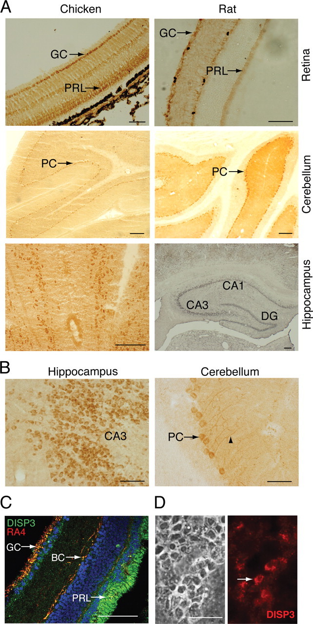
DISP3 is expressed in vertebrate neural cells. A, Representative micrographs of DISP3 immunoreactive cells in the chicken and rat retina, cerebellum, and hippocampus (brown staining). Specific regions of the hippocampus regions CA1, CA3, and dentate gyrus (DG) are shown. GC, Ganglion cells; PRL, photoreceptors; PC, Purkinje cells. B, Magnified images of A, highlighting individual DISP3-positive cells. C, Chicken retina sections double stained with an anti-DISP3 antibody (green) and the RA4 antibody (red) that identifies ganglion cells (GC) and bipolar cells (BC), respectively. 4′,6-Diamidino-2-phenylindole (blue) was used to stain nuclei. Yellow cells highlight areas of colocalization. D, Indirect immunofluorescence performed on primary chicken retina cultures using the purified polyclonal anti-DISP3 antibody. Scale bar, 100 μm for all panels.
