Fig. 5.

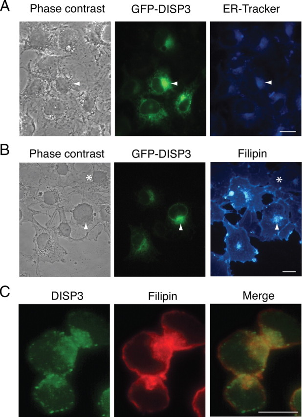
DISP3 localizes to the ER and colocalizes with cholesterol. COS7 cells transiently transfected with GFP-DISP3 were labeled with an ER-Tracker dye to show the sub-cellular localization of DISP3 (A) and by filipin to visualize cellular cholesterol (B). Arrowheads highlight selected DISP3-GFP-transfected cells; asterisks mark nontransfected cells. C, Y79 cells were stained with Disp3 antibody (green) and filipin (red-pseudocolored). Yellow staining represents areas of colocalization. Scale bar, 10 μm.
