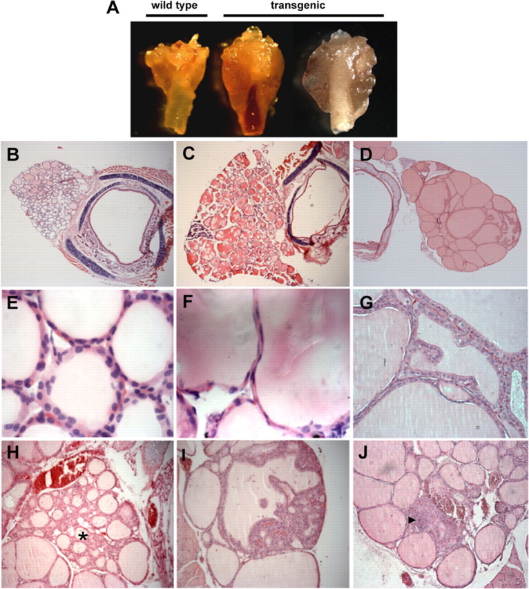
Fig. 2.

Thyroid histopathology in 9- to 12-month-old mice. A, Gross appearance of thyroid glands from wild-type (left) and transgenic mice with diffuse goiter (middle) or multinodular goiter (right). B–J, Hematoxylin and eosin staining of paraffin sections from wild-type (B and E) and transgenic (C, D, F–J) mice. C and F, Transgenic glands showing follicles with large lumens surrounded by flat epithelium. D and G, Hyperplastic areas with follicles surrounded by tall epithelium forming papillary infoldings protruding into the lumen. H, Adenomatous hyperproliferation (asterisk). Nodules with disorganized follicular structure (I) and nonfollicular hyperproliferative foci (arrowhead in J). Similar heterogeneity and pathological phenotypes were observed in both transgenic line LeuBTA7 (D, G, H, and J) and LeuBTA15 (A, C, F, and I). Magnification, ×50 (B–D), ×1000 (E and F), ×400 (G), and ×200 (H, I, and J).
