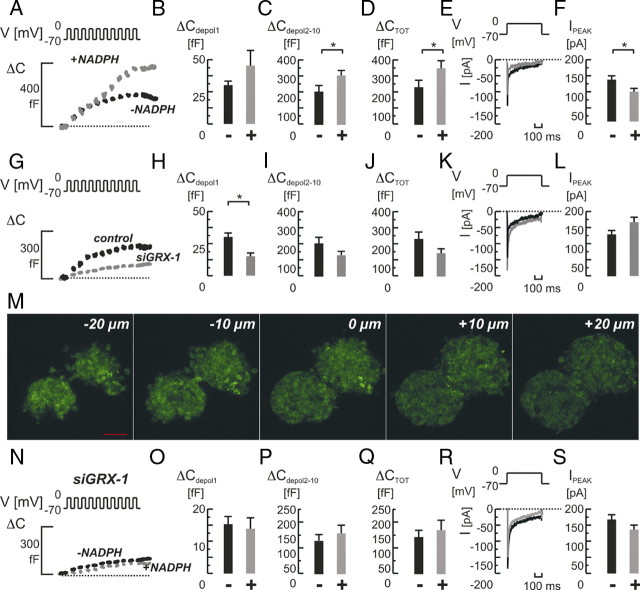
Fig. 5.
Role of GRX-1 knockdown for NADPH-mediated effects on single-cell exocytosis and whole-cell Ca2+ currents in primary rat β-cells. A, Depolarization-evoked (V) increases in cell capacitance (ΔC) in control cells with (+; in gray) or without (−; in black) NADPH (0.1 mm) added intracellularly. NADPH had a strong stimulatory effect on exocytosis (A). The effect on the response to the first pulse (ΔCdepol 1; panel B) was milder than on the response to later pulses (ΔCdepol2-10; panel C). Total exocytosis (ΔCTOT; panel D) was increased. E, Typical examples of whole-cell Ca2+ current recordings (E) in control cells with (in gray) or without (in black) NADPH added. F, Average peak Ca2+ currents (IPEAK) were significantly reduced. The knockdown of GRX-1 with siRNA (siGRX-1) reduced the exocytotic response significantly (G and J). The early response was unchanged (H), and especially the late response was affected (I). Whole-cell Ca2+ currents were similar (K and L). M, Confocal microscopy of pancreatic islets transfected with siGRX-1 and simultaneously a fluorescein-labeled dsRNA oligomer reveal the efficient uptake and distribution of siRNA throughout the islets (−20 to +20 μm in 10-μm steps from the islet center at 0 μm). The knockdown of GRX-1 now rendered NADPH an ineffective stimulator of exocytosis (N–Q). A mild inhibition on Ca2+ currents remained (R and S). Scale bar, 50 μm. Bar graphs represent average values ± sem. *, P < 0.05. ms, Milliseconds.

