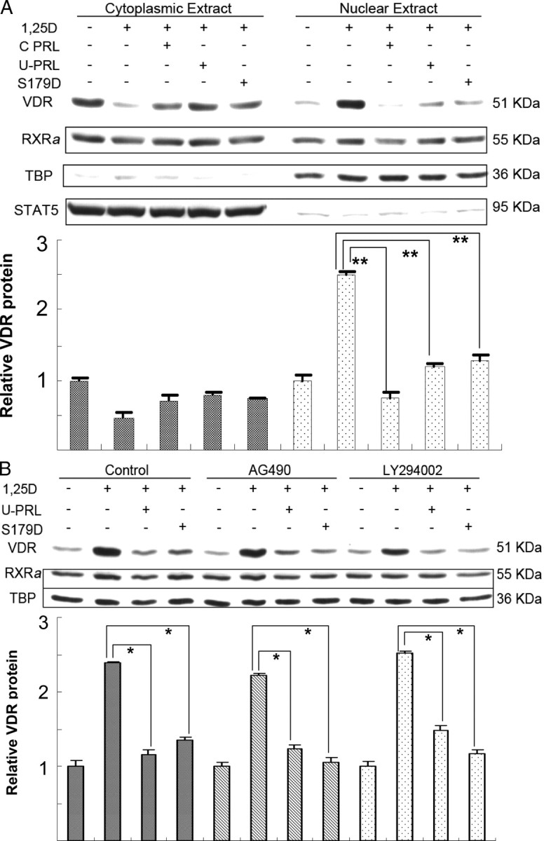
Fig. 5.

1α,25(OH)2D3-induced nuclear translocation of VDR is blocked by PRL. Ros 17/2.8 osteosarcoma cells were cultured in DMEM with 10% FBS and 1% PS and then incubated in the absence of FBS for 48 h before treatment with 1 nm 1α,25(OH)2D3 or this plus 200 ng/ml commercial recombinant hPRL (C PRL) or U-PRL or S179D PRL or diluent for 30 min. Subcellular fractionation and protein extraction were performed using NE-PER reagents. Ten percent of either the nuclear protein or cytosolic protein was loaded per lane. Western blots were performed using TBP as a marker of nuclear protein and total Stat5 as a marker of cytosolic protein. For panel B, cells were incubated in 50 μm AG490 or LY294002 for the last 2 h of the 48-h preincubation without FBS and then were processed as for panel A except that the inhibitors continued to be present, this time at 25 μm. Quantification from multiple experiments was normalized to cells without hormone treatment. RXRa, Retinoid X receptor α; other abbreviations as for previous figures. *, P < 0.03; **, P ≤ 0.01.
