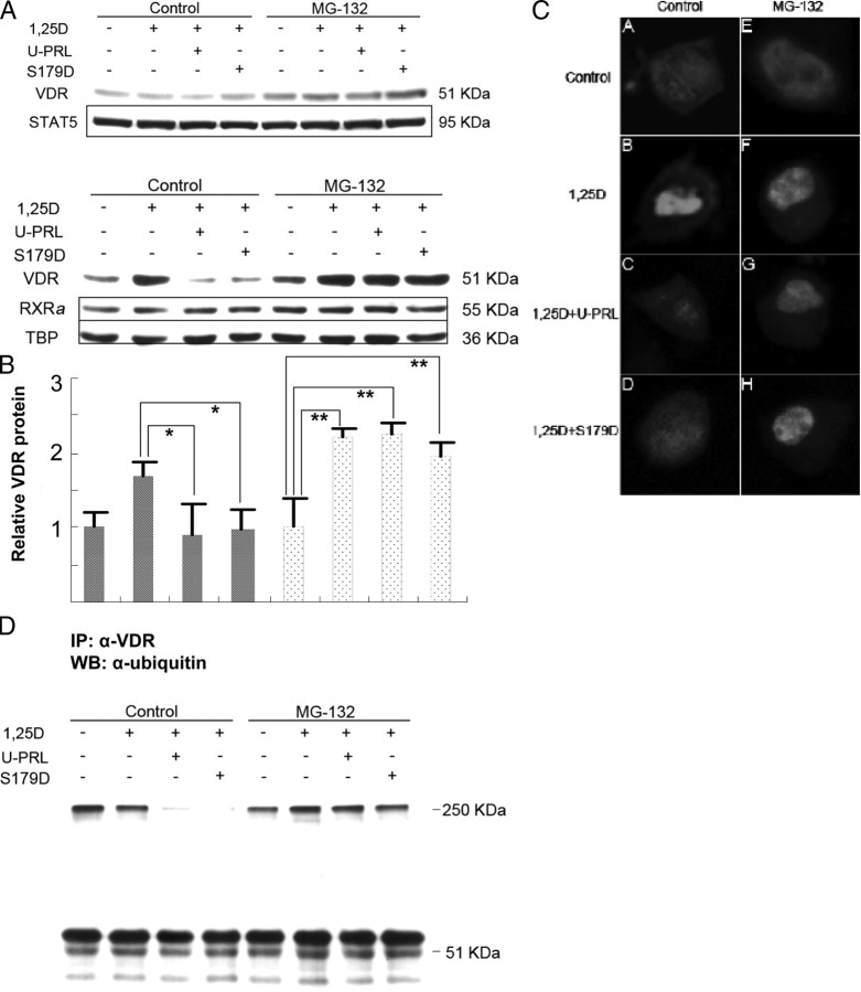
Fig. 6.
Short-term effect of MG-132 on the PRL inhibition of nuclear accumulation of the VDR. Ros 17/2.8 osteosarcoma cells were cultured in DMEM with 10% FBS and 1% PS and then incubated in the absence of FBS for 48 h. MG-132 (50 μm) was added 2 h before the end of this period. Cells were then incubated in 25 μm MG-132 and 1 nm 1α,25(OH)2D3 or this plus 200 ng/ml U-PRL or S179D PRL or diluent for 30 min. Whole-cell lysates are shown in panel A and nuclear extracts in panel B. Quantification from multiple experiments was normalized to cells without hormone treatment. For panel C, cells were transfected with pECFP-hVDR in the absence of FBS for 24 h and then incubated for a further 24 h in the absence of FBS. For the cells receiving MG-132, this was added as before: 2 h before the end of the total 48-h period in the absence of FBS and during the 30-min incubations. At the end of the 30-min incubations, cells were fixed in 4% formaldehyde in Dulbecco’s PBS at room temperature for 15 min, mounted in antifade, and observed by confocal fluorescence microscopy. The same samples as in panel A were used for the immunoprecipitation shown in panel D. From whole-cell lysate 500 μg protein per lane was used for immunoprecipitation with anti-VDR polyclonal antibody and then Western blotting with antiubiquitin monoclonal antibody. *, P < 0.05; **, P < 0.0004; 1,25D, 1α,25(OH)2D3; IP, immunoprecipitation; U-PRL, unmodified PRL; WB, Western blot.

