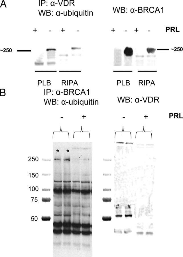
Fig. 7.
Identification of p250 as BRCA1. Ros 17/2.8 osteosarcoma cells were cultured and incubated in the absence of FBS as before and then treated with U-PRL or diluent for 30 min. Cells were extracted with either Promega passive lysis buffer (PLB) or radioimmune precipitation assay buffer, and the protein extract was then subjected to immunoprecipitation (IP) with either anti-VDR (α-VDR) for panel A or anti-BRCA1 for panel B. After transfer to nitrocellulose, Western blots with antiubiquitin were performed. Filters were then stripped, reblocked, and reprobed with either anti-BRCA1 (panel A) or anti-VDR (panel B). WB, Western blot.

