
Fig. 5.
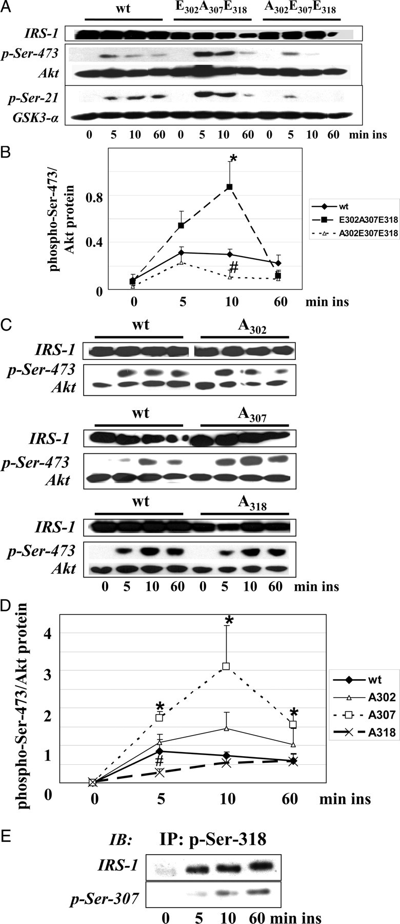
The Early Phosphorylation Pattern Enhances Insulin Action
A, C2C12 cells were transfected with IRS-1 wt, IRS-1 E302A307E318, or IRS-1 A302E307E318 and stimulated with 10 nm insulin as indicated. Cell extracts were immunoblotted for phospho-Ser-473 of Akt, phospho-Ser-9/21 of GSK-3, and IRS-1. Membranes were reprobed for Akt and GSK-3 protein. Only phosphorylation of Ser-21 of GSK-3α was detected, whereas both GSK-3α and -β were expressed (data not shown). B, Densitometric quantification of the phosphorylation of Ser-473 of Akt (n = 4, mean ± sem); *, P < 0.05 IRS-1 E302A307E318 vs. IRS-1 wt; #, P < 0.05 IRS-1 A302E307E318 vs. IRS-1 wt after 10 min of insulin stimulation. C, C2C12 cells were transfected with IRS-1 wt, IRS-1 A302, A307, or A318 and stimulated with 10 nm insulin as indicated. Cell extracts were immunoblotted for phospho-Ser-473 of Akt and IRS-1. Membranes were reprobed for Akt protein. D, Densitometric quantification of the phosphorylation of Ser-473 of Akt (n = 4, mean ± sem); *, P < 0.05 IRS-1 A307 vs. IRS-1 wt; #, P < 0.05 IRS-1 A318 vs. IRS-1 wt after 5, 10, or 60 min of insulin stimulation. E, Immunoprecipitation of IRS-1 wt transfected C2C12 cells using immunopurified anti-phospho-Ser-318 antibodies. Immunoprecipitates were immunoblotted for phospho-Ser-307 and IRS-1. IB, Immunoblot; ins, insulin; IP, immunoprecipitation.