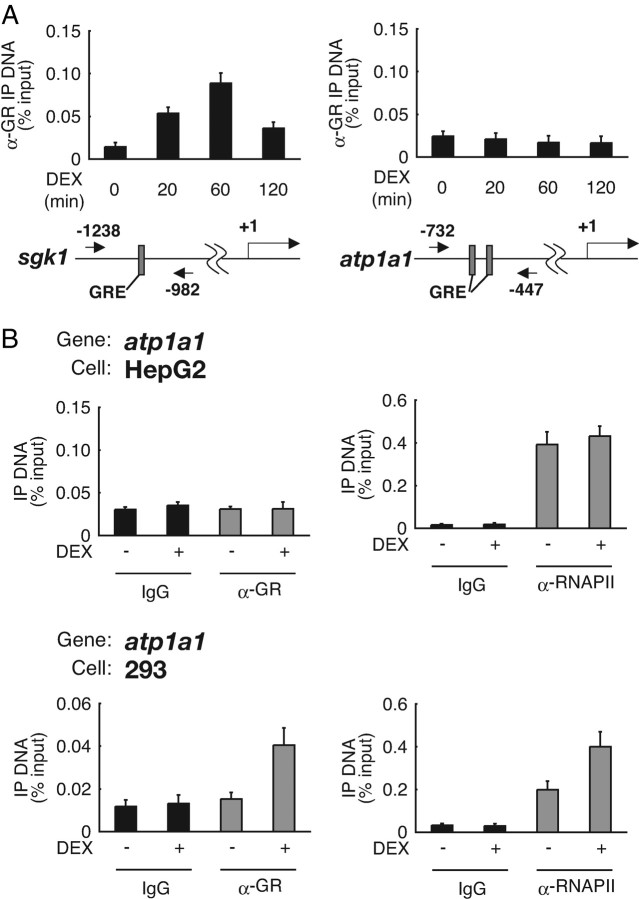
Fig. 2.
Cell Type Difference in Hormone-Dependent GR Recruitment onto atp1a1 Promoter GRE
A, HepG2 cells were cultured in phenol red-free Opti-MEM I for 24 h and treated with 1 μm DEX for the indicated time periods. ChIP assays were performed with anti-GR polyclonal antibodies, and recovered GRE-containing DNA fragments were measured with qRT-PCR. GREs in sgk1 and atp1a1 5′-flanking regions are indicated as gray boxes. The positions of the primers are shown as numbered arrows. Values are expressed as percentage of immunoprecipitated DNA to input. Error bars represent sd values of at least three independent experiments. B, HepG2 cells and 293 cells were cultured as described in panel A and treated with 1 μm DEX for 60 min. ChIP assays were performed with the indicated antibodies and primer sets as described in Materials and Methods. IP, Immunoprecipitation.

