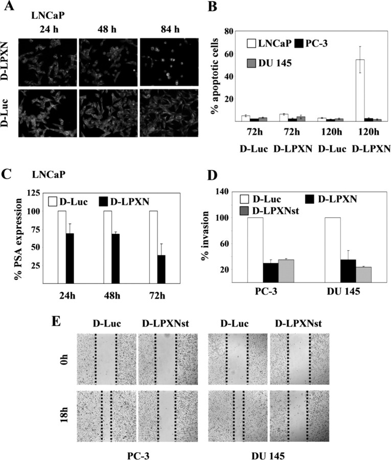
Fig. 7.
Down-Regulation of Leupaxin Expression Leads to Distinct Cellular Effects in Different PCa Cells
A, Androgen-dependent LNCaP cells with reduced leupaxin expression changed their morphology. The cells were grown on slides, transfected with leupaxin-specific (D-LPXN) and luciferase-specific (D-Luc) siRNA oligonucleotides, and incubated for the time periods as indicated. Subsequently, cells were fixed and stained with an anti-α-tubulin antibody to visualize the cell structure. Immunofluorescent images were acquired using ×100 magnification. B, Androgen-dependent LNCaP cells underwent spontaneous apoptosis 120 h after transfection with leupaxin-specific siRNA. LNCaP, PC-3, and DU 145 cells were transfected with leupaxin-specific (D-LPXN) and luciferase-specific (D-Luc) siRNA oligonucleotides. At 72 and 120 h after transfection, attached and floating cells were collected and cytocentrifuged on glass slides, and caspase 3 activities were detected as described in Materials and Methods. C, After leupaxin knockdown in LNCaP cells, subsequent inhibition of PSA secretion into the culture medium was detected at different time points (24, 48, and 72 h) using a PSA-specific ELISA. D, Androgen-independent and invasive PC-3 and DU 145 cells showed a reduction in their invasiveness in an in vitro Matrigel assay after down-regulation of leupaxin expression using leupaxin-specific siRNA (D-LPXN). Luciferase-siRNA-transfected cells were used as controls (D-Luc). E, Scratch assay analysis demonstrating a reduced direction migration of both PC-3 and DU 145 cells after transfection with siRNA against leupaxin (D-LPXNst) as compared with control-transfected cells (D-Luc).

